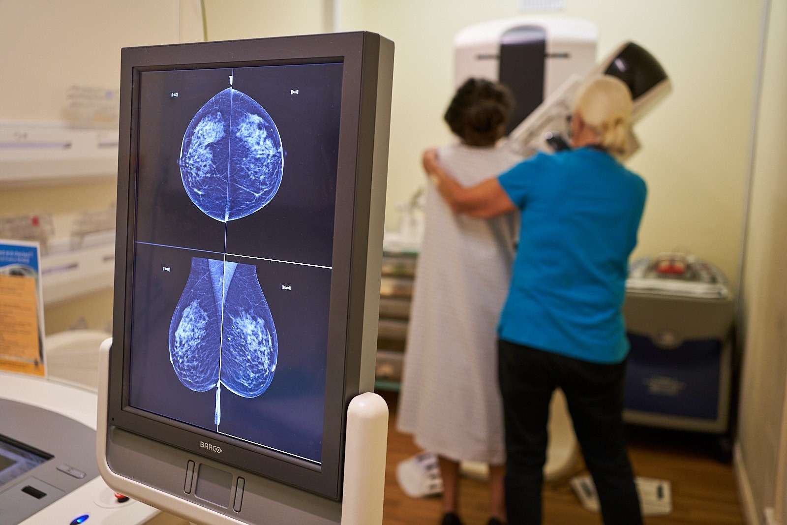
What happens during your appointment?
When you arrive, your mammographer will explain the procedure to you and will ask you to complete some paperwork to ensure that this test is suitable for you. You will have the opportunity to ask any questions you may have at this time.
During the examination, a contrast dye is injected into the arm and after waiting two minutes for the contrast to move through the body into the breast, two sets of mammograms are taken. Through specialist computer software, one image is subtracted from the other which leaves a contrast enhanced image highlighting any areas of interest, and providing detailed information to determine the position of the lesion within the breast.
Similar to a contrast enhanced MRI, breast lesions with an increased blood supply absorb more ‘dye’ than the surrounding breast tissue, making them more easily visible than a routine a mammogram.
The whole appointment will take around 30 minutes and if required, extra views can also be performed within this time.
Benefits
- Appointments are quick, taking around 30 minutes.
- In multiple published studies, Enhanced mammography has been known to have equal or near equal sensitivity to MRI but with a shorter procedure time and lower cost.
- Enhanced mammography has greater diagnostic accuracy than conventional mammography.
- Enhanced mammography can be performed in clinic as an immediate diagnostic tool rather than having to schedule an MRI, thus, speeding up the patient pathway and significantly reducing delays in diagnosis.
When would we use Enhanced Mammography?
- to assess patients with dense breast tissue, as breast density can increase the risk of breast cancer
- in cases where standard breast imaging (mammograms and ultrasound scans) may not be clear
- surveillance of high-risk patients; if there is a family history of breast cancer and particularly if there are known to be abnormal breast cancer genes in the family (such as BRCA1 and BRCA2)
- as an alternative for patients who cannot have MRI due to a pacemaker or other implanted medical device or those who suffer from claustrophobia
- to provide extra information about an existing breast cancer
- patients under 40 with breast malignancy
- assessment of patients with existing cancer to exclude further disease (this is called multi-focal breast cancer)
- in patients having chemotherapy, it can be a good way of monitoring treatment success
- a helpful tool in following up patients who have had breast cancer surgery
Not suitable
There are some cases when an Enhanced Mammography examination may not be suitable:
- Pregnancy or breastfeeding
- Allergic to Iodine
- Known kidney disease (including kidney transplant)
- Presence of Diabetes
- Currently taking Metformin
- Inability to tolerate mammogram because of physical problems
- Inability to give informed consent
Risks
- A very small number of people may be allergic to iodine, which is present in the contrast we use.
- In very rare cases, the dye used can also affect the kidneys.
Prior to your test, our clinical team will conduct a thorough risk assessment. This is to confirm no past allergic reactions while also checking for any risk factors.
Our consultant advice
Meet the team
Learn more about our team of consultants and the rest of your care team.


